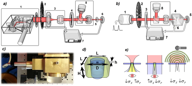
Figure 2.

(a) Schematic view of the experimental setup for AO measurements. Here we display the polarized pump source (1), the chopper (2), the optic group of lenses (3) used to shrink the light source, the QWP (4), the sample (5) and the photodetector (6). A lock-in (7) control the signals and a set composed of a beam splitter and a microscope (8) is adopted to visualize and correctly aim the helices. (b) Schematic view of the experimental setup for PA measurements. We show the laser source (1), the chopper (2), the QWP (3), the PA chamber (4) containing the studied sample and the PD (6); the microphone (6) is partially installed inside the PA chamber; here again a lock-in (7) is connected to the main set-up. (c) PA chamber with Mic attached on its left side. (d) Schematic view of the main part of the PA chamber. L = 6 cm, H = 3,5 cm, D = 4 cm, h = 1 mm. Quartz glasses insulates the air chamber from external space. (e) Illustrative representation of a near-field optical (left), far-field optical (middle), and photoacoustic (right) measurement set-up. The colors associated to distinct zones refer to specific field characters: red = laser source; yellow = transmitted light through the helices with a high incidence angle; green = transmitted light through helices with normal incidence (desired data); grey = mixed field arising both from helices’ region and nude substrate region; light grey = light purely transmitted by naked substrate. This representation simplifies the reasons behind the adoption of PA in the place of more common optical approach: if we want to observe the field emerging purely from helices (green), we must located the observer in close proximity of the helices, and use high-precision confocal systems; an excessive focusing will add unwanted plane components (high σ P) to the measured field (yellow); a far-field optical set-up is easier to use, but it rather detects a mixed field (high σ X) predominantly coming from the substrate (grey), and the resulting CD will be largely suppressed. The adoption of PA allows to detect the pure helices’ footprint without the need of sophisticated systems like that of first AO set-up, still achieving the same precision and reliability.
