Figure 3.

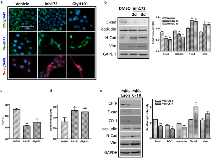
Suppression of CFTR induces EMT in MDCK cells. (a) Immunofluorescence staining showing decreased expression of ZO-1 and increased expression of N-cadherin and Vimentin in inh172 (10 µM for 2 or 3 days)- or GlyH101- (2.5 µM, 5 µM and 10 µM for 1 day) treated MDCK cells. Scale bar = 50 µm; (b) Western blot analysis showing increased expression of N-cadherin and Vimentin, and decreased expression of E-cadherin and Occludin in 10 µM inh172-treated MDCK cells. (Full-length blot is shown in Supplementary Figure S7e.) Quantification analysis is shown in the right panel, *p < 0.05, **p < 0.01; (c) TER (Transepithelial resistance) is significantly lower in CFTR inhibitor-treated MDCK cells. *p < 0.05, **p < 0.01, n = 3; (d) Wound healing assay showing enhanced cell migration at 24 hours with CFTR inhibitors treatment. At least 4 random fields per assay were counted and triplicate wells were set up for each sample. *p < 0.05; (e) Western blot showing decreased expression of ZO-1, E-cadherin and increased expression of N-cadherin and vimentin in CFTR knockdown cells. (Full-length blot is shown in Supplementary Figure S7f.) Quantification is shown in the right panel, *p < 0.05; **p < 0.01.
