
Figure 7.
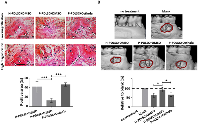
Osthole treated P-PDLSC sheet shows enhanced bone formation in animal model. (A) Representative images of H & E staining of H-PDLSC sheet, P-PDLSC sheet and P-PDLSC sheet with 10−7 Mol/L Osthole transplants in nude mice (arrow: new bone). Quantification of regenerated bone was calculated. Scale Bar, 50 μm (upper) and 100 μm (lower). (B) Micro CT analysis showed repair of bone defect in SD rats after transplantation of H-PDLSC sheet, P-PDLSC sheet and P-PDLSC sheet with 10−7 Mol/L Osthole. *P < 0.05, ***P < 0.001, n = 3.