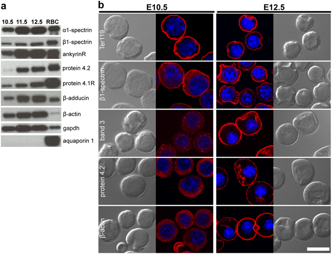
Figure 3.

Expression of membrane skeleton proteins in primary primitive erythroblasts. (a) Immunoblots of whole cell lysates prepared from 5 × 10⁵ cells loaded per lane. RBC, adult RBCs. Representative data from one of two or three independent experiments are shown. (b) Immunofluorescence of cytoskeletal proteins (red) in primitive erythroblasts from E10.5 (left panels) and E12.5 (right panels) mouse embryos. Parallel brightfield and fluorescence images are shown. Nuclei were stained with Hoechst (blue). Band 3, protein 4.2, and β-actin become localized to the plasma membrane only at late stages of maturation. Representative data from one of three independent experiments are shown. Scale bar represents 10 µm.
