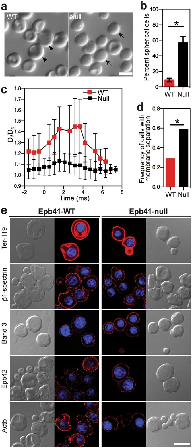
Figure 5.

Epb41-null primitive erythroblasts at the OrthoE stage (E12.5) have abnormal cell shape and defects in physical membrane features. (a) Morphology of freshly isolated Epb41-wild-type (WT) and Epb41-null primitive erythroblasts at E12.5. Spherical cells, arrows; partially concave cells, arrow heads. (b) The percentage of spherical cells at E12.5. Numbers represent at least 300 total cells per sample and error bars are SEM of three independent samples per stage. *p = 0.0028. (c) Aspect ratio (Dl/Ds) changes of deforming E12.5 Epb41-WT and Epb41-null primitive erythroid cells in the microfluidic channel. Dl, cell length; Ds, cell width. Time 0 indicates the time when cells enter the constriction channel. (d) AlexaFluor 488-Ter119 labeled E12.5 Epb41-WT and Epb41-null primitive erythroblasts were analyzed using fluorescence imaged deformation (FIMD). A significantly higher frequency of Epb41-null primitive OrthoE display evidence of membrane separation. *p = 0.0212. (e) Immunofluorescence of cytoskeletal proteins (red) in Epb41-WT and Epb41-null E12.5 primitive erythroblasts. Nuclei were stained with Hoechst (HO). Scale bar represents 10 µm.
