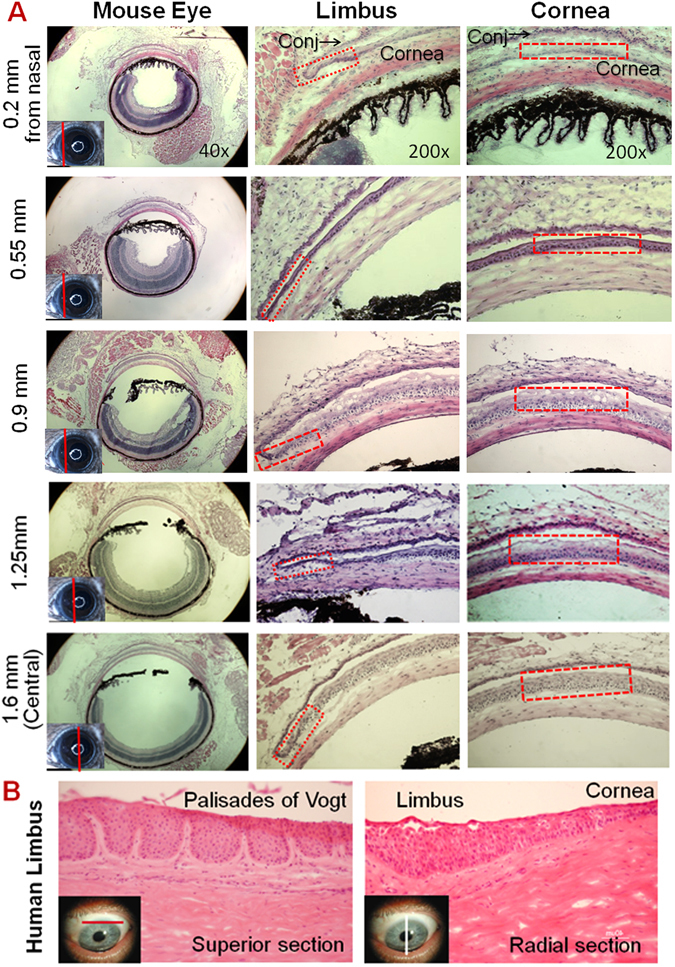
Figure 1.

Representative Haematoxylin-Eosin (H & E) staining images of mouse and human corneas. (A) Five sets of images were chosen from a series of mouse corneal cross sections from nasal to central cutting. The left column shows images of whole eye sections at different positions as red line on small eye inserts. The middle column shows limbal structures with red rectangles indicating the limbal epithelium containing 1~3 layers of cells. The right column shows corneal structures with red rectangles indicating the 5–7 layers of central corneal epithelium. (B) Human limbal sections with vertical or horizontal cutting show the palisades of Vogt structures.
