
Figure 4.
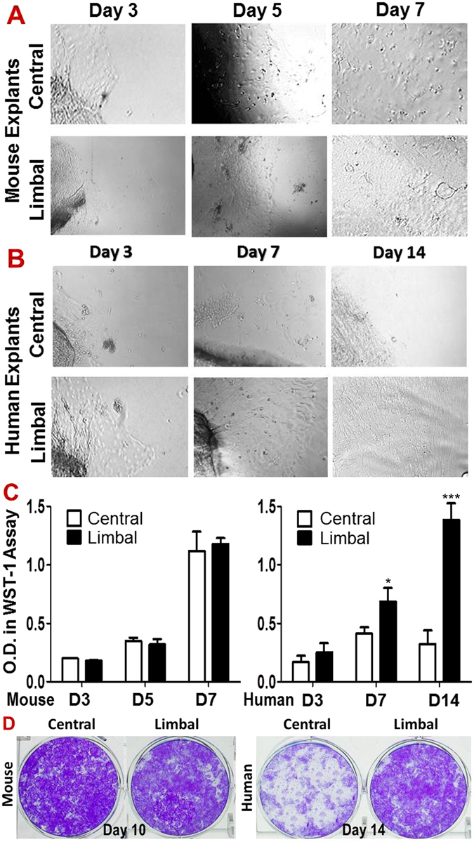
Comparison of proliferative capacity in primary corneal epithelial cultures between limbal and central cornea in mouse and human. (A) Phase-contrast microscopy showed the mouse epithelial cell growth of explants from limbal and central corneas on days 3, 5 and 7; (B) Human epithelial cell growth of explants from limbal and central corneas on days 3, 7 and 14. (C) WST-1 assay showed no difference of epithelial cell growth between limbal and central corneal explants from mice during 7 days (n = 5, P > 0.05); but there is growth difference between limbal and central corneal explants of human during 14 days. Data were summarized from 5 separated experiments. *P < 0.05 and ***P < 0.001, as two groups compared. (D) The representative images from clonogenic assays of the single cells from corneal or limbal epithelia in mice and human.