Figure 5.

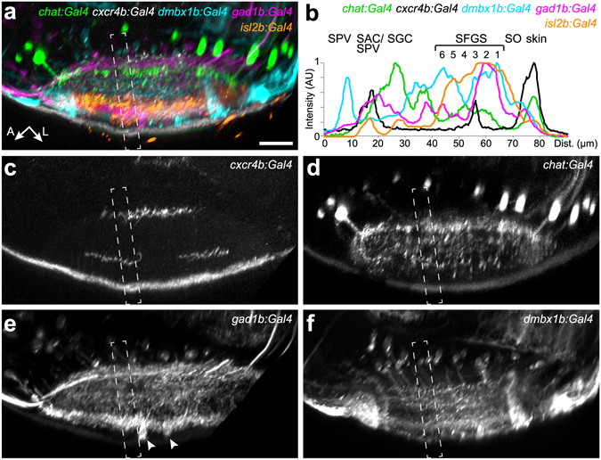
New transgenic lines label distinct sublaminae in the tectal neuropil. Lateral view of the tectal neuropil shows registered expression pattern of chat:Gal4, cxcr4b:Gal4, dmbx1b:Gal4, gad1b:Gal4, and isl2b:Gal4, as merged (a) or single channels (c–f). (b) Fluorescence intensity plots along the boxed regions in (a–f). Intensity peaks of isl2b:Gal4 expression were used for layer determination. SIN cell bodies labeled by gad1b:Gal4 are marked by arrowheads in (e). The peak for dmbx1b:Gal4 in the SPV layer reflects labeled periventricular cell bodies.
