
Figure 2.
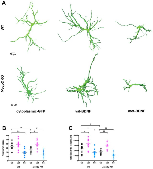
val-BDNF increases, and met-BDNF reduces dendritic length and branching in both Mecp2 KO and WT neurons, without affecting axonal morphology. (A) Representative 3D reconstructions of WT and Mecp2 KO hippocampal neurons expressing either soluble GFP alone, val-BDNF and soluble GFP, or met-BDNF and soluble GFP. Note that these are images from anti-GFP immunofluorescence labeling, and they reveal the full morphology of the neurons (the GFP tags of val-BDNF and met-BDNF are masked by soluble GFP, which fills the entire cell). (B) Number of dendritic branch points (nodes). (C) Total dendritic length, identified as MAP2(+) processes. *p < 0.05, **p < 0.01, compared to WT; #p < 0.05, ##p < 0.01 compared to Mecp2 KO.