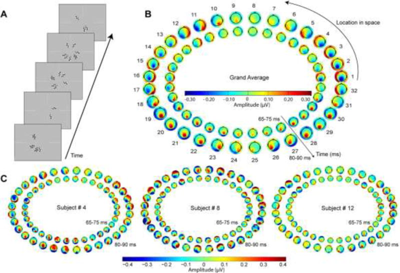
Figure 1. Data from preliminary individualized mapping procedure.

A) Shown are randomly chosen screen captures during the pattern-pulse multifocal stimulation. B) The grand average scalp potential topographies across all 15 subjects of the earliest component of the pattern-pulse VEP for each of the 32 stimulus locations, in the time frames of 65–75 ms (inner) and 80–90 ms (outer). The typical average topographic shifts consistent with calcarine cortical physiology (see Kelly et al. 2013b) are readily observable, with remarkable symmetry about the vertical meridian. Segments 18 and 31, for example, correspond to the fundus of the calcarine sulcus in the right hemisphere for lower left visual field stimulation (18) and in the left hemisphere for lower right visual field stimulation (31), respectively (Clark et al. 1995). C) Topographies of three individual subjects selected to highlight the inter-subject variability in topographic dependence on location, consistent with the well-known variability in calcarine cortical size and geometry (Amunts et al. 2000; Rademacher et al. 1993). Whereas Subject 4 (left) exhibited typical dipolar field shifts as a function of polar angle with vertical symmetry, Subjects 8 and 12 exhibited maps that clearly diverged from the average pattern in terms of points of apparent dipole rotation and asymmetry. For each individual, a single right-hemifield polar angle was identified at which a strong, approximately midline-focused, negative C1 was evoked, which was then used in the main experiment manipulating stimulus contrast and size. Polar angle of the thus chosen segments ranged from −11.25° to 45° (mean: 18.00°; SD: ±14.13°) relative to the right horizontal meridian.
