Fig. 9.
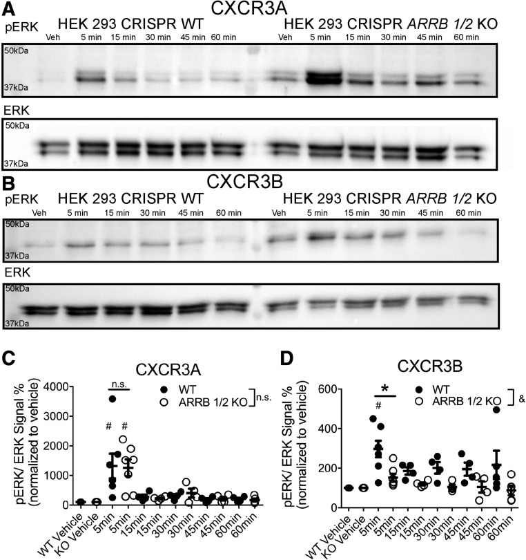
β-arrestin KO differentially alters pERK signaling between CXCR3 splice variants. WT or ∆ARRB1/2 cells transfected with CXCR3A (A) or CXCR3B (B) and stimulated with CXCL11 (100 nM). A significant increase in pERK1/2 signal was observed in both WT and ∆ARRB1/2 cells transfected with CXCR3A. However, only WT, but not ∆ARRB1/2, cells transfected with CXCR3B displayed a significant increase in pERK1/2 signal. The CXCR3A (C) and CXCR3B (D) phospo-ERK signal was quantified in WT and ∆ARRB1/2 cells [note the change in scale of the y-axis between (C) and (D)]. In (C) and (D), &P < 0.05 indicates a significant effect of cell line by two-way analysis of variance (ANOVA) in cells transfected with CXCR3B; *P < 0.05 indicates a significant difference in the 5-minute time point by two-way ANOVA followed by Bonferroni post hoc comparison (corrected for all time points) in cells transfected with CXCR3B; #P < 0.05 by one-way ANOVA and Dunnett’s post hoc comparison of CXCL11 stimulation to the respective isoform vehicle control at the indicated time point; ± S.E.M., n ≥ 3 biologic replicates per condition. Western blots shown are representative of at least three separate experiments.

