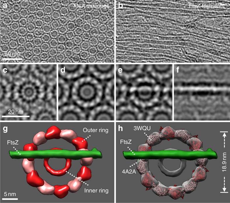
Figure 5. Tomography and structural modelling of FtsZ protofilaments aligned on top of FtsA minirings on lipid monolayers.
(a,b) Two tomographic slices of the same transmission electron microscopy (TEM field), from the lipid monolayer upward, showing a FtsA miniring hexagonal array (a), then aligned unbundled FtsZ protofilaments on top of the array (b) (see also Supplementary Movie 1). (c–f) A subvolume-averaged portion of a field from (a,b), with tomographic slices from the lipid monolayer (c) up to FtsZ filaments (f). (g) Segmentation of the 3D reconstructions showing potential sites of interaction where an FtsZ filament (green) intersects with the FtsA miniring (red and pink subunits). (h) Atomic structures from S. aureus FtsA (3WQU) are shown docked into the miniring densities, along with the FtsA-binding peptide from the C terminus of FtsZ (4A2A) (green) into the two subunits interacting with the crossing FtsZ protofilament. See also Supplementary Movie 2. For simplicity, the other 10 FtsZ C-terminal peptides are not shown.

