Figure 1.
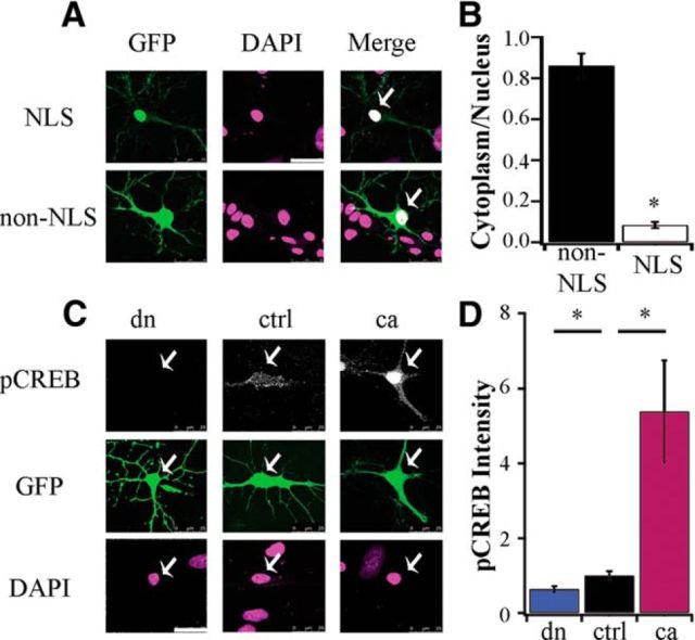
Cell-autonomous, bidirectional manipulation of nuclear CaMKIV signaling. A, Example pyramidal neurons transfected for 24 h with NLS tagged construct (top) or soluble GFP without NLS (bottom). The fluorescent signal of GFP (green) and DAPI (magenta) are shown. Scale bar, 25 μm; white arrows indicate nucleus. B, Quantification of the cytoplasmic-to-nuclear fluorescent intensity ratio for NLS and non-NLS-expressing neurons (GFP n = 8, 0.86 ± 0.06; NLS n = 9, 0.09 ± 0.02; t test, p < 0.001). C, Example pyramidal neurons transfected for 24 h with dnCaMKIV (dn), control (ctrl) or caCaMKIV (ca), along with soluble GFP to visualize cell morphology; fixed and stained with an antibody against endogenous phospho-CREB Ser133 (grayscale). The fluorescent signal of GFP (green) and DAPI (magenta) are also shown. D, Quantification of the nuclear fluorescent intensity of phospho-CREB Ser133 (normalized to ctrl; dn, n = 20, 0.64 ± 0.08; ctrl, n = 47, 1 ± 0. 11; ca, n = 27, 5.39 ± 1.37; *p < 0.05, ANOVA followed by post hoc Games–Howell test).

