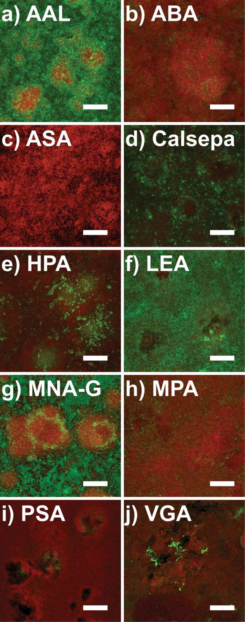
Figure 1.

Confocal laser scanning microscopy (CLSM) images of 48 h biofilms grown in situ stained with fluorescently labeled lectins. Panels a–j show maximum intensity projections of biofilm images from one subject, with the FITC-labeled lectins in green and the nucleic acid stain SYTO® 60 in red. Aleuria aurantia (AAL), Calystega sepiem (Calsepa), Lycopersicon esculentum (LEA), and Morniga-G (MNA-G) show strong fluorescence signals. Helix pomatia (HPA) shows strong selective binding inside dense bacterial clusters. Vicia graminea (VGA) binds selectively to bacterial surfaces in branched spider web-like colonies. Agaricus bisporus (ABA), Allium sativum (ASA), Maclura pomifera (MPA), and Pisum sativum (PSA) show rather diffuse or no signals at all. Scale bars = 25 µm.
