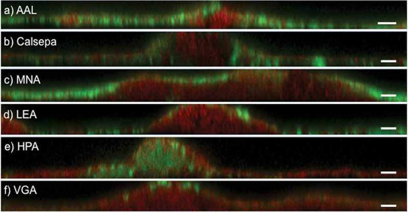
Figure 2.

Deconvoluted CLSM images of 48 h biofilms grown in situ stained with fluorescently labeled lectins. Panels a–f show representative xz projections of lectin-stained biofilms (green). AAL, Calsepa, LEA, and MNA-G bind on the biofilm surface and in thin areas of the biofilms. HPA binds selectively to dense cell clusters in protuberations of the biofilms. VGA binds selectively to the surface of some organisms. Bacterial staining was performed with SYTO® 60 (red). Scale bars = 5 µm.
