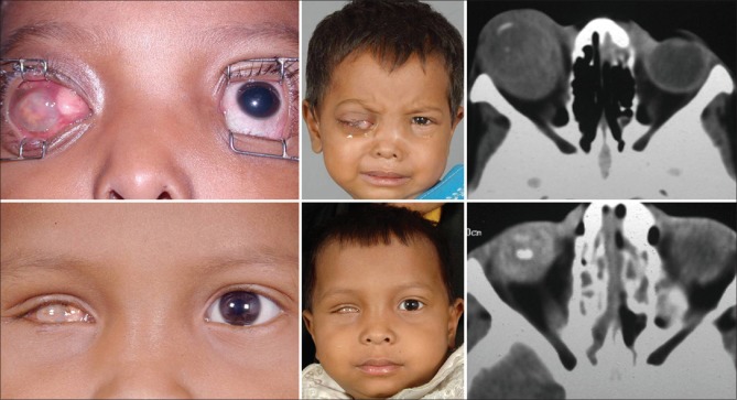
Figure 9.
Outcome of management of primary orbital retinoblastoma. A 3-year-old child with primary orbital retinoblastoma in the right eye with an anterior episcleral nodule (top left and middle), with axial computed tomography scan showing massive orbital extension (top right). Following 3 cycles of high-dose neoadjuvant chemotherapy, the right eye has undergone a quiet phthisis (bottom left and middle), with the computed tomography scan shows complete resolution of the orbital tumor (bottom right)

