
Figure 2.
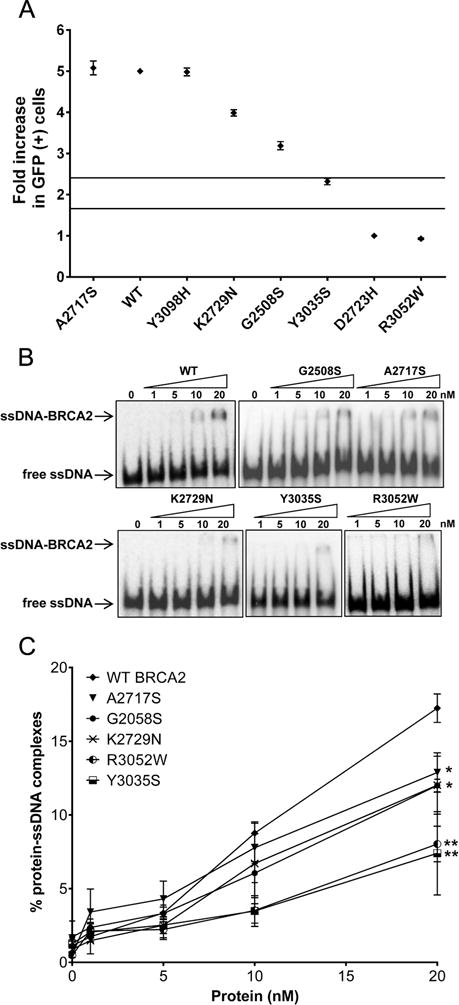
(A) HDR and ssDNA binding activity of BRCA2 p.Y3035S is reduced. (A) Activity of BRCA2 missense variants is shown as HDR fold change with standard error (SE) (of three independent measures of duplicates) on a scale of one to five. Solid lines represent 99.9% and 0.1% probability of pathogenicity. (B) Representative Electrophoretic Mobility Shift Assays (EMSA) of DNA-protein complexes formed by mixing increasing concentrations (0, 5, 10, 20 nM) of purified BRCA2 wildtype and mutant proteins with ssDNA. (C) Quantitation of the DNA-protein complex formation shown in Fig. 2B. Error bars represent SE derived from at least three independent experiments. Statistical difference between WT and mutant BRCA2 protein-DNA complexes formation was determined by two-sample t-test. **p<0.001; *p<0.05. WT, wildtype.