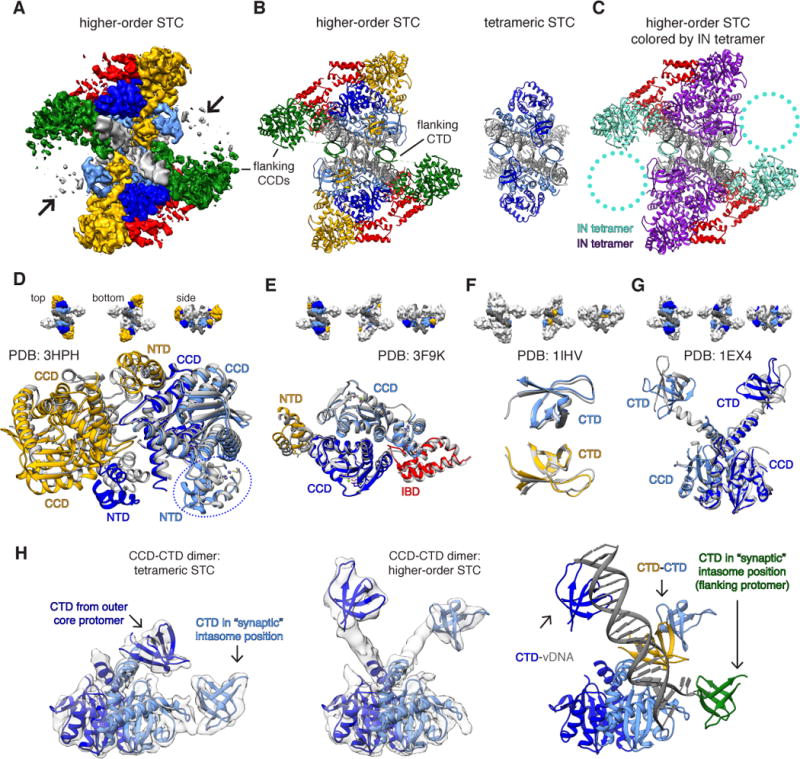
Fig. 3. HIV-1 STC intasomes form higher order oligomers through distinct mechanisms of assembly.

(A) CryoEM density map of IBD-bound STCs (STCIBD). Densities are segmented either by IN protomer (inner core: light blue, outer core: dark blue) or IN dimer (yellow and green), while IBD is in red. (B) Higher-order STC model assembled by rigid-body docking individual domain components, colored as in A. The higher-order STC (left) is shown side-by-side with the tetrameric STC from Fig. 1 (right). (C) Model as in B, colored by IN tetramer (28). Circled regions contain poorly resolved density that may harbor additional IN dimers. (D–G) Structural comparison of higher-order STC assembled through rigid-body docking of individual domains with prior multi-domain IN structures. The structural components of higher-order STCs are colored as in panels A–B, while the PDB structures used for comparison are gray. Comparisons include: (D) MVV INNTD-CCD tetramer (PDB: 3HPH, IBD has been omitted for clarity; the circled NTD arises from an IN protomer on the opposite side of vDNA), (E) HIV-2 INNTD-CCD dimer bound to IBD (PDB: 3F9K), (F) HIV-1 CTD dimer (PDB: 1IHV), and (G) HIV-1 INCCD-CTD dimer (PDB: 1EX4). (H) Conformational rearrangement within the core CCD-CTD dimer between (left) the tetrameric STC and (center) a higher-order STC, both overlaid on respective filtered experimental EM density. At right, the rearranged higher-order dimer is displayed in the context of additional CTDs and vDNA within the asymmetric unit. The “synaptic” position is required to form the conserved intasome core interface present in all retroviral intasomes.
