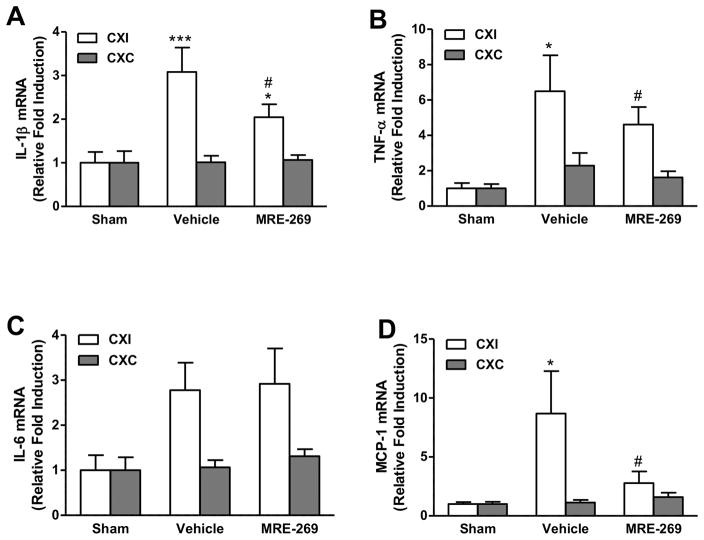
Figure 3. Effect of MRE-269 on cortical gene expression of cytokines and chemokines in aged rats following transient MCAO.
Aged male rats underwent 90 min of MCAO and were randomly selected to receive either vehicle or MRE-269 (0.25 mg/kg; i.v.) given at 4.5 h and an additional dose at 12 h after stroke onset. Animals were sacrificed 18 h post-MCAO and perfused with ice-cold saline, the ipsilateral and contralateral cerebral cortex were saved for RNA extraction and western blot studies. Transcript levels of IL-1β (A), TNF-α (B), IL-6 (C), and MCP-1 (D) were determined by RT-qPCR. Data were expressed as mean ± SEM, *P<0.05, ***P<0.001 versus sham and #P<0.05 versus vehicle. Sham, n=5; Vehicle, n=12; MRE-269, n=13. CXI = cortex ipsilateral to stroke; CXC = cortex contralateral to stroke.

