
Figure 1.
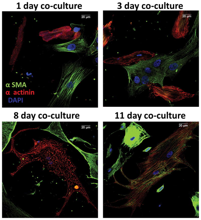
Evolution of cardiomyocyte trans-differentiation in co-culture with myofibroblasts. Fluorescence labeling for α-smooth muscle actin (α-sma, green), α-actinin (red) and DAPI (blue). At one day in co-culture, most cardiomyocytes express α-actinin and are rod-shaped and striated. At three days, many myocytes are no-longer rod shaped and acquire neonatal/fetal phenotype. On day 11, surviving myocytes beat spontaneously, are 4–5x larger than normal, show filopodia and stain strongly for α-sma in addition to α-actinin. Right, negative control. Calibration, 20 μm in all frames.