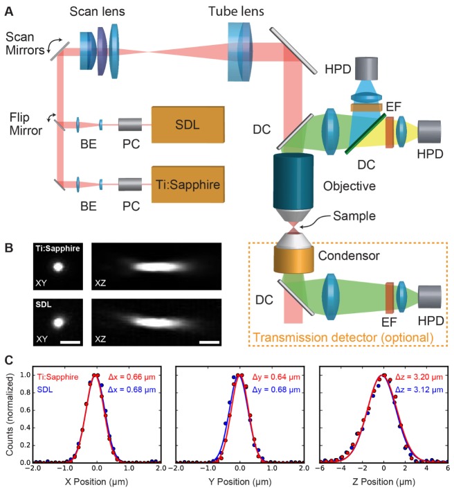
Fig. 4.
A) Overview of the microscope setup: Excitation light from either the SDL or a Ti:Sapphire laser (Spectra-Physics Mai Tai DeepSee) can be coupled into the microscope via a Pockels cell (PC) and a beam expander (BE). After the scan mirrors, the beam is directed into the objective by a scan and tube lens. The emission light is sent to a two-channel detection system via dichroic mirrors (DCs), emission filters (EF) and then collected by hybrid photo-detectors (HPDs). For small, transparent samples, a transmission detector with a single HPD can be added. B) Maximum intensity projections of images of the same 200 nm bead taken with the Ti:Sapphire and SDL. C) Gaussian fit of the data in B) with full width at half maximum (FWHM) values for both lasers.

