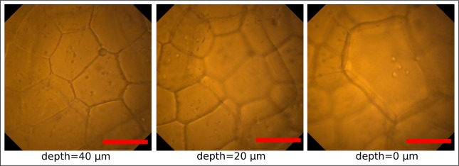
Fig. 5.
Pictures of the upper surface of a leaf of Begonia x ricinifolia captured with the endomicroscope at the same lateral position but at different axial distances. Probe has been attached to a five-axis stage and moved mechanically away from the specimen in 20 μm steps (from left to right). The ability to sharply image the different axial tissue layers consisting of different cell sizes with a uniformly high resolution but slight intensity reduction for deeper layers is observed. Scale bars 50 μm, respectively.

