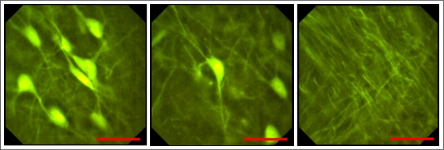
Fig. 6.
Pictures captured with the endomicroscopic probe of a thin histological brain slice of a transgenic Thy1-GFP-M mouse expressing enhanced green fluorescent protein in a sparse subset of neuronal populations. Illumination in epi-direction. The submicrometer resolution enables the visualization of cellular compartments like dendrites and somas. Scale bars 50 μm, respectively.

