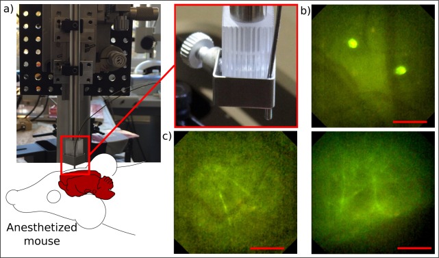
Fig. 9.
In-vivo imaging of neuronal cells through a cranial window implanted in the skull of an anesthetized Thy1-GFP-M mouse expressing green fluorescent protein in a sparse subset of neurons. a) Mouse has been placed on a heated stage. The endomicroscopic probe has been moved precisely by a five-axis-stage in lateral and axial direction to sharply image the region of interest. b) Visualization of dendritic cells and blood vessels directly below the cranial window. c) Measurements at up to 60 μm deep inside layer I of the neocortex reveals single dendrites and blood vessels. Scale bars 50 μm, respectively.

