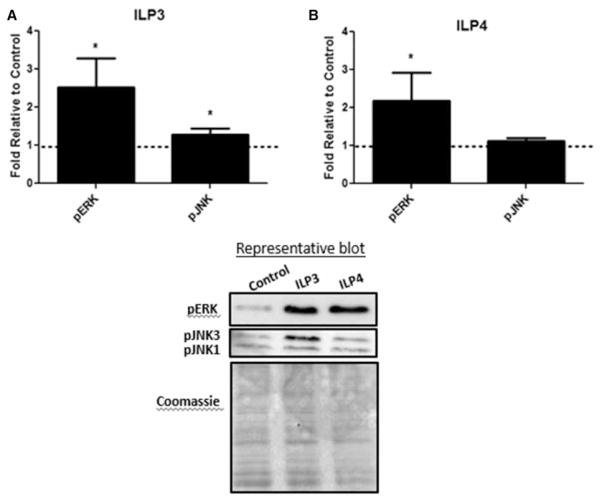
Figure 3. ILP3 and ILP4 signaling in the A. stephensi midgut.
Cohorts of A. stephensi were fed (A) 170 pM ILP3 or (B) 170 pM ILP4 in a meal of uninfected human RBCs. Midguts were collected 15 min after feeding and protein extracts were used for Western blotting to detect protein phosphorylation. Values from ILP-treated samples were normalized first to Coomassie brilliant blue stain for total protein and then to untreated controls (unsupplemented human RBCs; set at 1) for calculation of fold change. Experiments were independently replicated with 3–5 cohorts of 60–90 mosquitoes each and data were analyzed by t-test to determine differences between ILP-treated and control groups. *P-values were deemed significant when P ≤ 0.05.

