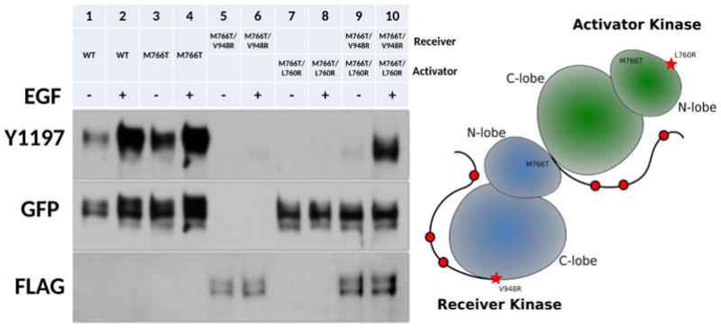
Figure 3.

Dimerization dependency of M766T mutation. Right panel: Lanes from left to right: WT EGFR (−), WT EGFR (+), M766T (−), M766T (+), M766T/V948R (−), M766T/V948R (+), M766T/L760R (−), M766T/L760R (+), M766T/V948R+M766T/L760R (−), M766T/V948R +M766T/L760R (+). − and + indicate the absence and presence of EGF stimulation. Left panel: a cartoon representation of EGFR asymmetric dimer with N-lobe dimerization deficient mutation (L760R) and C-lobe dimerization deficient mutation (V948R).
