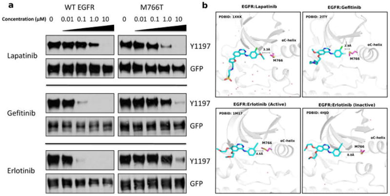
Figure 7.

(a) Drug sensitivity of WT and mutant (M766T) EGFR. Drug concentration from left to right: 0 μM, 0.01 μM, 0.1 μM, 1.0 μM, and 10.0 μM. Upper panel (lapatinib),44 middle panel (gefitinib),19 lower panel (erlotinib).43–45 (b) Structural binding mode of EGFR M766 with lapatinib/gefitinib/erloti nib.
