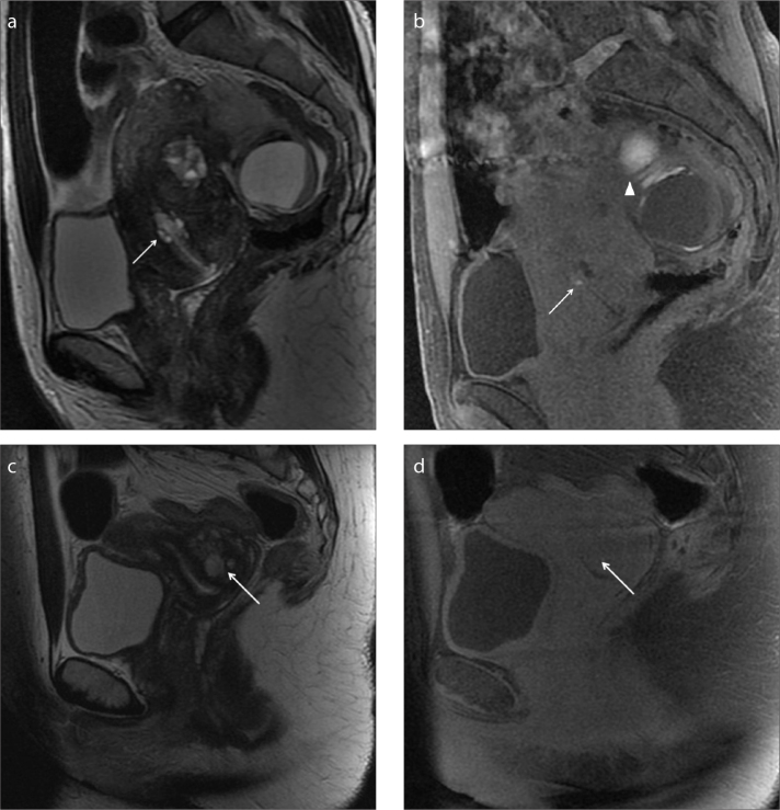
Figure 1. a–d.
A 45-year-old woman complaining of cyclic abdominal pain and spotting (a, b) and a second case of a 40-year-old woman complaining of spotting (c, d). Sagittal T2-weighted image (a) and T1-weighted image with fat-suppression (b) show a small bright area in the cervix, within the cervical stroma, hyperintense in both sequences as hemorrhagic content (white arrow). The patient has concomitant multiple signs of endometriosis on the left ovary (b, white arrowhead). In the second case, the sagittal T2-weighted image (c) and T1-weighted image with fat-suppression (d) show a focal area in the cervical stroma that appears hyperintense on T2-weighted images and isointense on T1-weighted images as fluid continent (c, d, white arrow). In spite of the MRI appearance, cervical biopsy reveals endometriotic hyperplasia.

