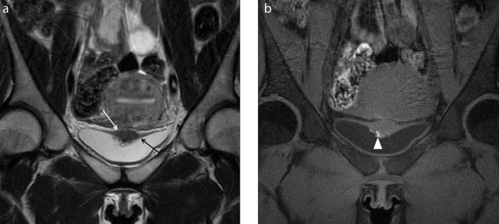
Figure 5. a, b.
A 30-year-old woman complaining of cyclic pelvic pain and discomfort during urination. Coronal T2-weighted (a) image shows focal and irregular hypointense wall thickening of the upper aspect of the bladder wall (white arrow) that obliterates the vesicouterine fold and distorts the bladder wall. The detrusor muscle seems to be infiltrated and the lesion seems to project into the lumen (black arrow). Coronal T1-weighted images with fat-suppression (b) show hyperintense foci within the mass that can be attributed to hemorrhagic content (white arrowhead). Laparotomy confirmed endometriotic localization of the vesicouterine fold with bladder wall involvement.

