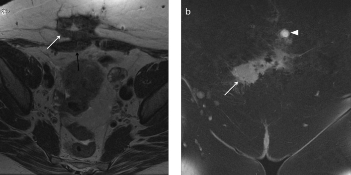
Figure 8. a, b.
A 45-year-old woman with history of previous cesarean sections suffering from localized abdominal pain at the site of a palpable parietal mass. Axial T2-weighted image (a) shows the presence of a hypointense spiculated mass in the subcutaneous soft tissue of abdominal wall (white arrow). The mass is strictly adjacent to the right rectus abdominis muscle (black arrow). The coronal T1-weighted images with fat-suppression (b) show slight hyperintense signal of the mass (white arrow) with some hyperintense areas due to hemorrhagic component; the coronal image reveals another small nodular lesion (white arrowhead) depicted cranially as hyperintense signal on T1-weighted image with fat-suppression. The lesions are due to abdominal wall deep endometriosis implants. Histopathology confirmed the endometriotic nature of the lesions.

