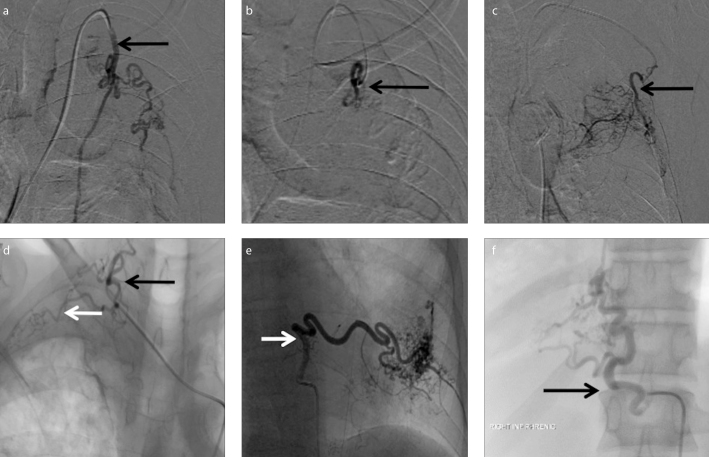
Figure 4. a–f.
Nonbronchial systemic collaterals. Preembolization DSA image (a) shows selective catheterization of hypertrophied left internal mammary artery (arrow) arising from left subclavian artery. Postembolization DSA image (b) shows successful selective embolization of left internal mammary artery branches with decreased vascularity and parenchymal blush (arrow). DSA image (c) of another patient shows selective catheterization of hypertrophied left lateral thoracic artery (arrow) arising from left subclavian artery. DSA image (d) shows hypertrophied right costocervical artery with normal cervical component (black arrow) and abnormal parenchymal blush from the costal component (white arrow) in another patient. DSA image (e) of a different patient shows selective catheterization of hypertrophied left posterior intercostal artery (arrow) with significant parenchymal blush. DSA image (f) of another patient shows hypertrophied right inferior phrenic artery (arrow).

