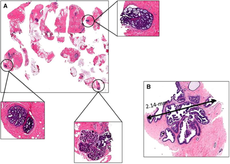
Figure 1.

Histological features predicting upgrade. A, multiple cores involved with atypical ductal hyperplasia (ADH), figure with scanning magnification shows three foci of ADH (in circles) with corresponding figures with higher magnification (in boxes); B, ADH focus measuring >2 mm (2.14 mm), the excision shows low nuclear grade ductal carcinoma in situ.
