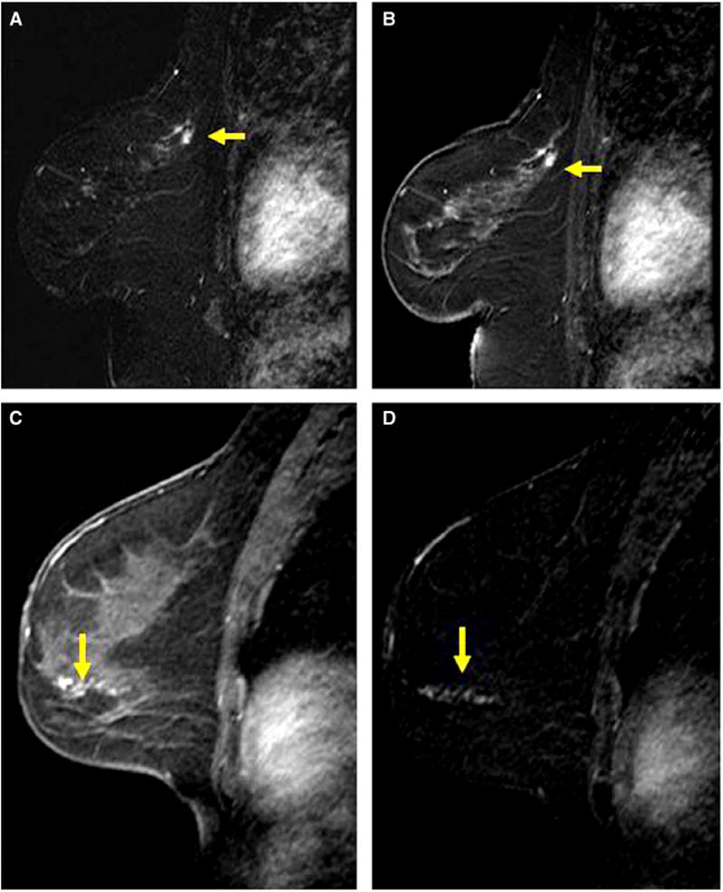
Figure 2.

Examples of mass and non-mass enhancement. A, B, Sagittal post-contrast magnetic resonance (MR) image (A) and corresponding subtraction image (B) of the left breast in a 59-year-old woman demonstrates a 0.8-cm irregular mass in the posterior superior breast (yellow arrows). Magnetic resonance imaging (MRI)-guided biopsy yielded atypical ductal hyperplasia (ADH) and excision yielded ADH and sclerosingadenosis. C, D, sagittal post-contrast MR image (C) and corresponding subtracted image (D) of the left breast in a 40-year-old woman demonstrate 4 cm of non-mass enhancement in a linear distribution (arrows). MRI-guided core biopsy yielded ADH and excision yielded low to intermediate grade ductal carcinoma in situ.
