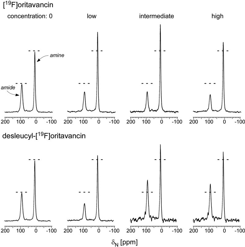
Figure 3.
(Top) 15N full-echo NMR spectra of S. aureus grown in media containing [1-13C]glycine and L-[ε-15N]lysine in the presence of varying concentrations of [19F]oritavancin (low, 3.4 µg/ml; intermediate, 6.7 µg/ml; high, 10.1 µg/ml). The spectra were scaled using the normalization factors of Figure 2 (left). Spectra of S. aureus grown in the presence of [19F]oritavancin show a reduced ε-15N lysyl-amide peak at 95 ppm (cell wall) and an increased ε-15N lysyl-amine peak at 10 ppm (cytoplasmic peptidoglycan precursors), consistent with transglycosylase inhibition. (Bottom) 15N full-echo NMR spectra of S. aureus grown in media containing [1-13C]glycine and L-[ε-15N]lysine in the presence of varying concentrations of desleucyl-[19F]oritavancin (low, 3.5 µg/ml; intermediate, 6.9 µg/ml; high, 10.4 µg/ml). The spectra were scaled using the normalization factors of Figure 2 (right). At low drug concentrations, the level of transglycosylase inhibition by desleucyl-[19F]oritavancin is reduced relative to that by [19F]oritavancin (lower amine-to-amide ratio). Slowed growths (Figure S2, top right) suggest the possibility of some accumulation of cell-wall debris from dead cells. Chemical shifts are referenced to solid ammonium sulfate.

