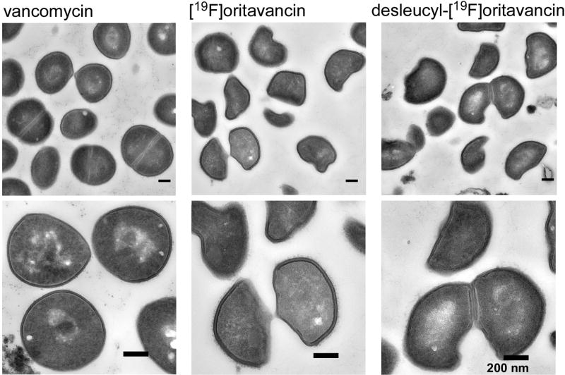
Figure 5.
Transmission electron micrographs of S. aureus treated with vancomycin (left column), [19F]oritavancin (middle column), and desleucyl-[19F]oritavancin (right column). The top row shows a representative, large field of view. Vancomycin-treated S. aureus shows no significant change in cell shape, but general thinning of cell wall with diffused cell-wall staining for newly formed cell walls at the plane of cell division. Oritavancin- and desleucyl-[19F]oritavancin-treated S. aureus show thinning of cell wall at the septum of dividing cells and an aberrant cell shape.

