Figure 1.

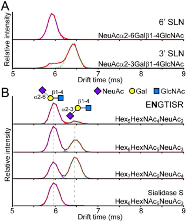
(A) Separation of α2-3 and α2-6 linked sialic acid by IMS. Arrival time distributions of the [M − H2O + H]+ fragment (m/z 657.24) of 3′ (top) and 6′ (bottom) sialyl-N-acetyllactosamine. The signal at ∼6 ms in the 3′ standard was not due to impurities (Figure S1) but most likely alternative gas phase conformations of the 3′ SLN standard. (B) Glycopeptide ENGTISR (residues 84–90) from a tryptic digest of α1AGP was isolated, subjected to collision induced dissociation, and the fragments were resolved by IMS. Differences in the ratio of α2-3 and α2-6 sialylation are evident between the biantennary bisialo (top), triantennary trisialo (2nd trace), and tetraantennary tetrasialo (3rd trace) glycoforms. Sialidase S treatment to specifically remove only α2-3 sialylation reveals an ATD showing purely the signal for the α2-6 linked species (bottom).
