
Figure 1.
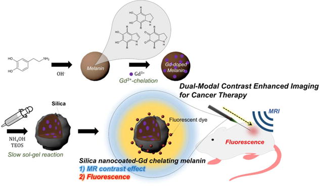
Illustration of gadolinium chelated melanin nanoparticles with silica nanocoating (Gd-Mel@SiO2 NPs) in preparation and application. Dopamine molecules start polymerization in the presence of hydroxide ions and end up making spherical melanin nanoparticles (Mel NP). Paramagnetic Gd3+ ions are chelated in melanin matrices for MRI T1 contrast effect. Then, a silica layer is formed on the surfaces of Mel NP with a slow sol–gel reaction controlling a feeding of precursor to reduce self-nucleation of free silica particles. Lastly, fluorescent dye molecules are labeled for the fluorescent imaging of Gd-Mel@SiO2 NPs without significant electron quenching. Our developed fluorescent dye-labeled Gd-Mel@SiO2 NPs are applied to dual-modal contrast enhanced MRI/fluorescent image-guidance for cancer therapy.