
Figure 5.
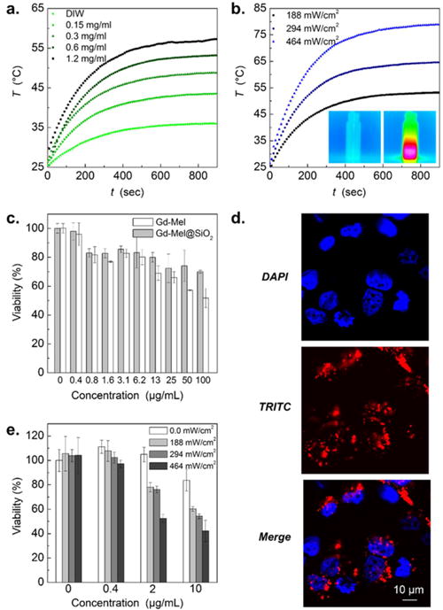
(a) Time-dependent increase of temperature in solutions of Gd-Mel@SiO2 NPs with concentrations of 0, 0.15, 0.3, 0.6, and 1.2 mg/mL, respectively, during near-infrared (NIR) radiation (188 mW/cm2). (b) Time dependent temperature changes of Gd-Mel@SiO2 NPs solutions with a concentration of 0.3 mg/mL, irradiated with three different powers of 808 nm NIR light (188 mW/cm2, 294 mW/cm2, and 464 mW/cm2), respectively. Insets of temperature mapping images indicate before and after NIR radiation, respectively. (c) Cytotoxicity of TRITC-labeled Gd-Mel NPs and TRITC-labeled Gd-Mel@SiO2 NPs against human prostate cancer cells (PC-3) with respect to the concentration. (d) Fluorescent images of PC-3 cells, stained with DAPI for nuclei and encapsulating TRITC-labeled Gd-Mel@SiO2 NPs, and the superposition of DAPI and TRITC, as indicated in each panel. (e) Photoheat treated cell viability depending on the concentration of TRITC-labeled Gd-Mel@SiO2 NPs (0, 0.4, 2, and 10 μg/mL) and power of NIR light (0, 188, 294, and 464 mW/cm2).