
Fig. 5.
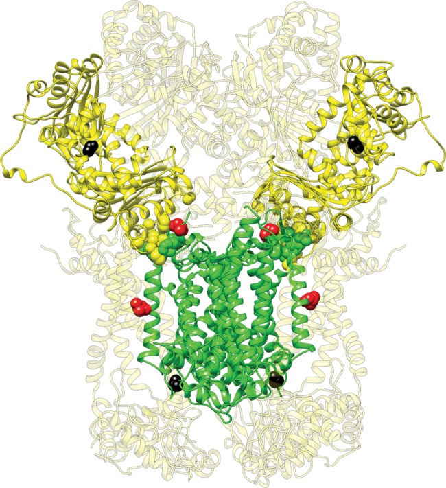
—The fixed amino acid substitution between modern humans and Denisovans in the N-mt protein UQCRC1 mapped onto the solved crystal structure for cytochrome bc1. The mitochondrially encoded CYTB subunits (two monomers) are shown in green and the UQCRC1 subunits (two monomers) are shown in bright yellow. The other N-mt subunits are shown in light yellow. Variable residues and those that form the interface between CYTB and UQCRC1 are shown as spheres. Substitution at contact residues are shown in red, while substitutions at noncontact residues (including the one in UQCRC1) are shown in black.