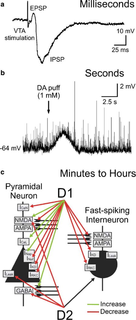
Fig. 1.

Orders of magnitude in the observed time course following dopamine (DA) application or VTA stimulation. a A very fast EPSP–IPSP sequence can be recorded in prefrontal cortical cells after stimulation of the VTA in-vivo. The EPSP is evoked with a latency on the order of milliseconds and is thought to be the result of corelease of glutamate from dopamine cells in the VTA. b Depolarization of a fast-spiking interneuron by DA in the prefrontal cortex in vitro. Local pressure application of DA leads to depolarization and repolarization of the membrane potential that seems to follow the diffusion of the drug in the slice on the timescale of seconds (Kroener and Seamans, unpublished observations). c Modulation of a variety of intrinsic and synaptic currents by DA has been shown to occur over minutes and hours both in vivo and in vitro. Activation of D1- and D2-type receptors occurs in both pyramidal cells and interneurons, adding to DA’s ability to modulate network behavior. The time course and direction of some of the effects indicated in the diagram have been shown to be concentration- and receptor-specific. It is assumed that in vivo the very long lasting effects that have been reported in experimental settings can be curtailed by fluctuating levels of extracellular DA and opposing effects at the different DA receptors that result from it. See text for details
