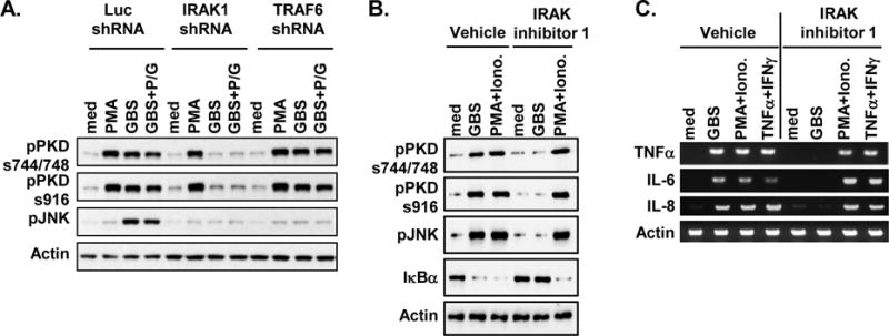
Figure 3. Activation of PKD1 induced by GBS is dependent on IRAK1, but independent of TRAF6.

(A) Control luciferase-knockdown (Luc-shRNA), IRAK1-knockdown (IRAK1-shRNA) or TRAF6-knockdown (TRAF6-shRNA) RAW264.7 cells were stimulated with medium, PMA (10 ng/ml), live GBS (108 cfu/ml), or antibiotic-killed GBS (GBS+P/G; 108 cfu of GBS were treated with 1 mg of penicillin G for 6 h) for 45 min. (B, C) THP1 cells were pretreated with vehicle (0.5% v/v DMSO) or IRAK inhibitor 1 (5 μM) for 1 hr and then stimulated with medium, GBS (1 × 10⁸ cfu/ml), or PMA (10 ng/ml) plus Ionomycin (10 ng/ml) for 1 hr (B) or 4 hr (C). Phosphorylation status of PKD (pPKDs744/748, pPKDs916) and JNK (pJNK), and protein levels of IκBα were detected by Western blot assay (A, B). Total RNA was isolated and mRNA levels of the indicated cytokines were analyzed by RT-PCR (C). Actin was used as a loading control. Data represent results obtained from three separate experiments.
