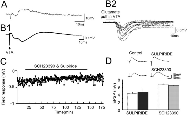
Figure 1.
Properties of the response in the PFC to single-pulse electrical stimulation of the VTA. A, Intracellular recordings from PFC neurons revealed an EPSP evoked by VTA stimulation. B1, Averaged traces from 32 field recordings in the PFC elicited by VTA stimulation revealed a multiphasic waveform. B2, Representative field responses from 13 animals in which a glutamate pulse (0.25-1 mm, 50 ms) was delivered to the VTA. C, The D1 and D2 receptor antagonists SCH23390 (10 μm) and sulpiride (10 μm) applied together to the PFC via reverse microdialysis did not modulate the field response significantly. D, Top, Representative traces in control and after sulpiride or SCH23390 application. Bottom, Group data showing that D1 (▪; 1 mg/kg SCH23390; n = 3) or D2 (⊡1.5 mg/kg sulpiride; n = 3) antagonists did not affect the mean amplitude of the VTA-PFCEPSP relative to control (□) recorded intracellularly, suggesting that the response is not DA mediated.

