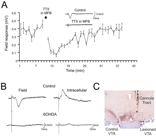
Figure 3.
Response in the PFC to VTA stimulation is blocked by MFB inactivation or 6-OHDA VTA lesions. A, A concentration of 1 μm TTX puffed to the MFB abolishes the averaged evoked amplitude of the postsynaptic component of the field response (n = 8). Inset, Representative trace of the field response before and after the TTX puff into the MFB. The stimulus artifact was cropped for clarity. B, Left, Field response in the PFC evoked by VTA stimulation in a control rat (top) and in a rat with a 6-OHDA lesion of the VTA (bottom). Right, Intracellular recording of a PFC neuron after VTA stimulation in a control rat (top) and from a rat with a 6-OHDA lesion of the VTA (bottom). C, Photomicrograph of a representative section through the VTA stained for TH.

