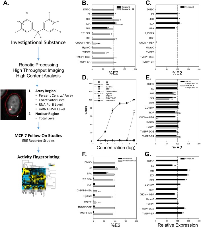
Fig 2. PRL-HeLa analysis of estrogenic and anti-estrogenic activity of investigational chemical compounds on ERα.
(A) Experimental workflow for analysis of investigational molecules. Percentage of cells with an array in GFP-ERα:PRL-HeLa cells after 1 hour of treatment with maximal dose of compound alone or with 10 nM E2 after 1 hour (B) or 24 hours (C) of exposure. Antagonism experiments were done combining 10nM E2 with 5 μM of the compounds of interest. (D) Dose-response curves of percent of cells with arrays for E2, BPA and the seven investigational compounds. (E) Effects on SRC-3 coactivator and RNA polymerase II levels at visible arrays after 1 hour of treatment with 10 nM E2 in the presence of maximal dose of compound. (F) Effects on de novo mRNA production at the PRL array after 1 hour of treatment with compound alone or with 10 nM E2. (G) Effects on GFP-ERα expression level after 24 hours of treatment with maximal dose of compound. All data represent mean of a minimum of 4 experiments ± standard deviation. (*) or (**) indicates a response significantly (p < 0.05) above or below control treatment.

