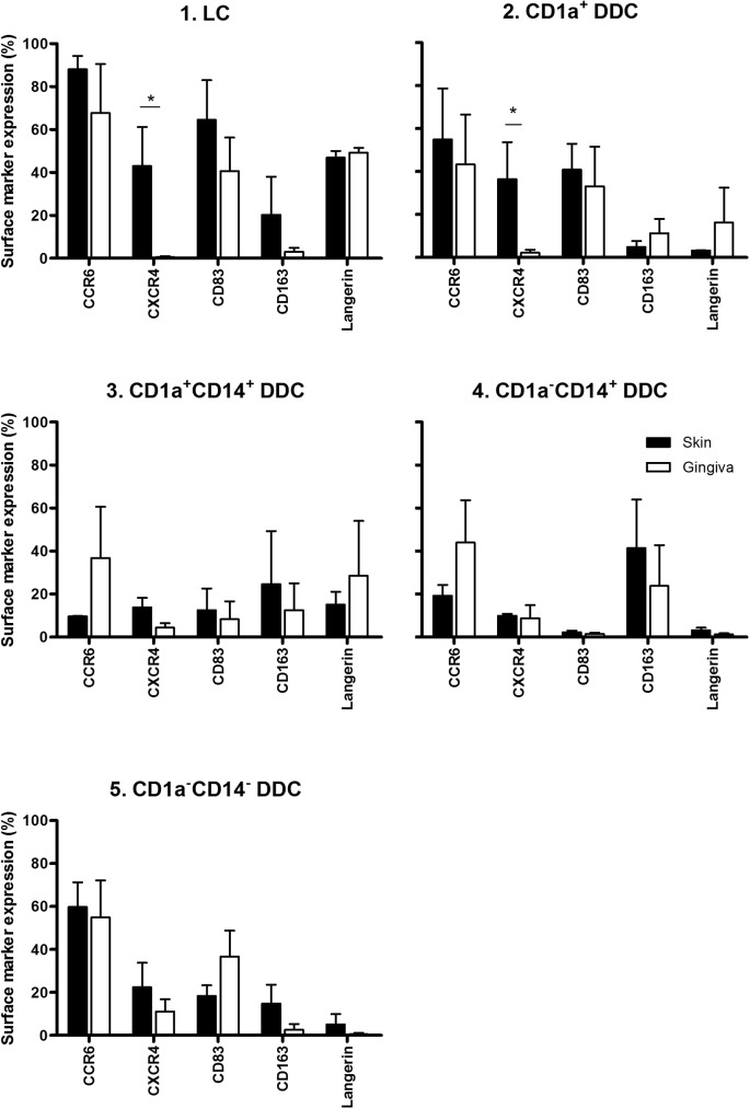
Fig 3. Phenotypic analysis of migratory dendritic cell (DC) subsets from skin and gingiva.
Chemotaxis, maturation and differentiation-associated marker expression on DC subsets 1–5 from skin vs. gingiva, shown per indicated subset (*P<0.05, n = 4–9 for skin and gingiva). Explants (6mm diameter) from skin and gingiva were taken and cultured floating in medium for 48h, after which they were discarded and migrated DC harvested, stained and analysed by flowcytometry.

