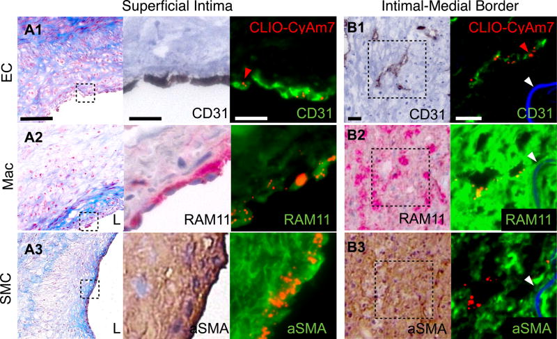
Figure 2. Cellular distribution of CLIO-CyAm7 nanoparticles in atherosclerosis.
CLIO-CyAm7 uptake by plaque endothelial cells (ECs), macrophages (Macs) and smooth muscle cells (SMCs). Carstairs’ stain shows structural characteristics of atheroma, including collagen (blue). (A) Carstairs’, immunohistochemical (IHC), and immunofluorescence (IF) stains for CD31 (A1), RAM11 (A2), and alpha-smooth muscle actin (aSMA, A3) show correspondence between CLIO-CyAm7 signal (indicated by red arrows) and superficial, luminal ECs, macrophages, and SMCs, respectively. (B) Distribution of CLIO-CyAm7 along the intimal-medial border in regions of neovasculature as detected by CD31 stain (B1). CLIO-CyAm7 deposition occurred in areas of RAM11+ macrophages flanking the neovasculature (B2). IHC: RAM11+ = red; CD31+ and aSMA+ = brown. FM fusion images: CLIO-CyAm7 = red, IF antibodies = green, autofluorescence = blue. White arrows = internal elastic membrane. Scale bars: Low magnification Carstairs’ = 100µm, IF and IHC = 25µm. L=vessel lumen.

