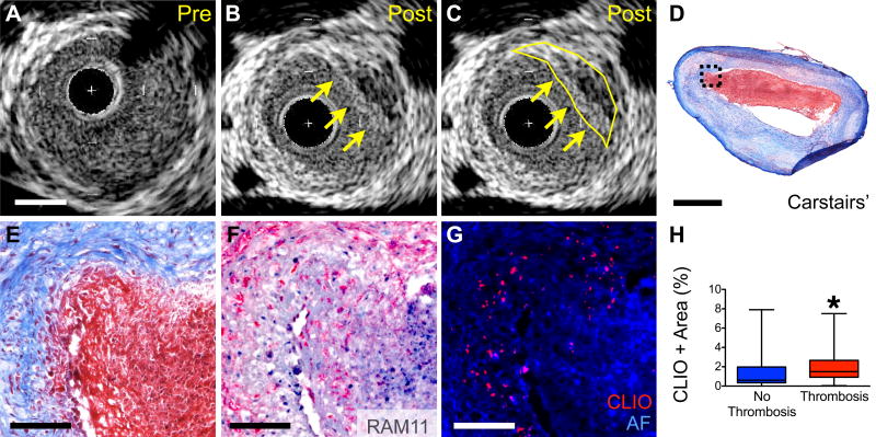
Figure 5. In vivo and microscopic analyses of nanoparticle deposition and triggered plaque thrombosis.
(A) Cross sectional IVUS image of rabbit aorta with atheroma prior to pharmacologic triggering. (B,C) Post-triggering IVUS image corresponding to (A) demonstrating new luminal irregularity (yellow arrows and outline) consistent with new thrombus (segmented in (C)). (D,E) Corresponding histology of plaque with adherent thrombus. Carstairs’ staining of fibrin rich adherent thrombus (red). (F) RAM11+ macrophages are present at the surface below the thrombus. (G) Epifluorescence microscopy revealing increased CLIO-CyAm7 (red) at the plaque shoulder and underlying areas of thrombus. (H) Significantly higher CLIO-CyAm7 accumulation occurred in regions with thrombosis compared to atheroma without thrombosis (2.1±1.7%, n=34, and 1.5±1.9%, n=23, respectively, p=0.045). Scale bars: IVUS and low magnification histology = 1mm, high magnification histology = 100µm.

