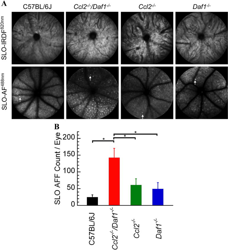
Fig. 1.
SLO imaging of mouse models studied. (A) Representative SLO images obtained from C57BL/6J, Ccl2−/−/Daf1−/−, Ccl2−/−, or Daf1−/− mice. Top panels show no appreciable morphologic differences across genotypes for SLO-IRDF imaging at 820 nm. Bottom panels use SLO-AF imaging at 488 nm, which shows the (AFF) in mutant strains compared to C57BL/6J. A single AFF is indicated with an arrow in each SLO-AF488 nm image. (B) Numbers of SLO-AF AFF. Bars indicate the mean ± SE for 5–8 mice per genotype. The number of SLO-AF AFF is significantly higher in Ccl2−/−/Daf1−/− in comparison to all genotypes (all p < 0.05); the other comparisons are not significant.

