
Figure 4. SDS-PAGE Analysis of Crude C. l. klauberi (C.L.K) and C. l. morulus (C.L.M) Venoms and Anion Exchange Protein Groups (I, II, III, IV, V, VI) of C. l. morulus Venom.
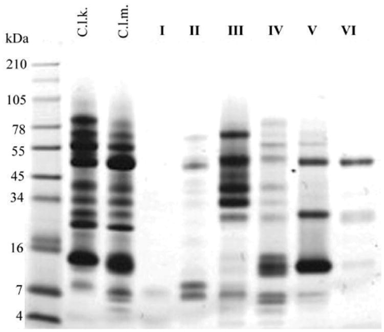
A total of 20 μg of crude venom and 10 μg of venom fractions were separated under reducing-conditions in a 10-20% Tricine acrylamide gel. The gel was run at 120V for 90 min using a BioRad PowerPak system. Proteins were stained with Simply Blue Safe Stain (Invitrogen). Numbers on the left and right of the gel indicated migration of the molecular mass markers.