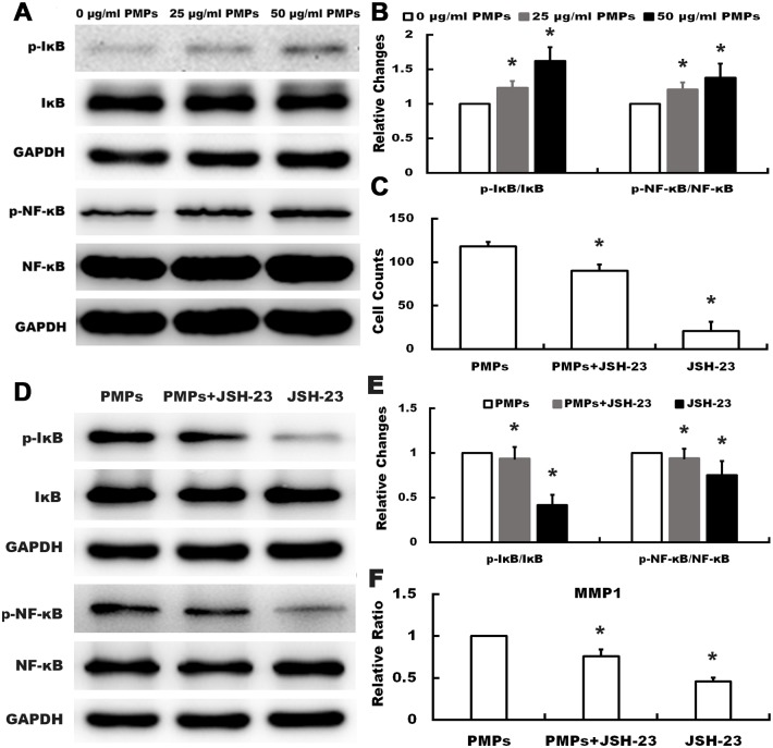
Fig 5. Effects of PMPs on the expression and activation of IκB and NF-κB in RA-FLSs.
(A and B) Western blotting was performed to detect the expression of p-IκB, IκB, NF-κB and p-NF-κB of RA-FLSs after treated with PMPs. The quantification was expressed as fold-change of 0μg/ml PMPs. (*p < 0.05 vs. 0 μg/ml PMPs) (C) Quantification of the migration assay of RA-FLSs treated with PMPs and JSH-23 was shown. (*p < 0.05 vs. PMPs) (D and E) After treated with PMPs and JSH-23, the expression of p-IκB, IκB, p-NF-κB and NF-κB of RA-FLSs were detected by western blot. (*p < 0.05 vs. PMPs) (F) After treated with PMPs and JSH-23, the expression of MMP1 was detected by RT-qPCR. (*p < 0.05 vs. PMPs).

